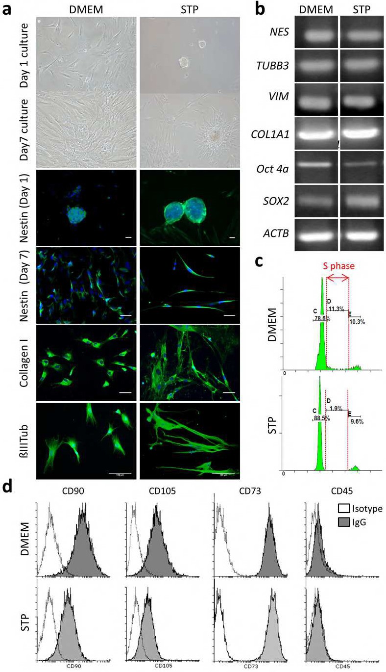
Fig. 1. Characterization of hDPSCs cultures in DMEM+10% FBS and STP media: a) Different growth patterns depending on the culture medium. Migrating adherent cells soon colonize the whole plating surface in DMEM+10% FBS. In contrast, floating dentospheres persist in STP medium, with occasional emergence of long spindle-like adherent cells. IF; images showing expression of mesenchymal markers (Collagen I) and neural markers (Nestin, β3-Tubulin) in hDPSCs in both DMEM+10% FBS and STP culture media. Scale Bar 50µm. b) Conventional RT-PCR derived electrophoresis gel bands showing the expression of mesenchymal genes, neural genes and pluripotency core factor genes in both conditions. c) Cell cycle analysis by propidium iodide flow cytometry of hDPSCs grown in both culture media, showing a limitation of the growth rate in STP medium, with respect to DMEM+10% FBS. d) Flow cytometry analysis for mesenchymal and hematopoietic stem cell markers (CD90, CD105, CD73, CD45) confirmed a generalized ectomesenchymal phenotype of hDPSCs in both media conditions.